Home
/ Knee Tendon Diagram : Knee Ligaments and Other Knee Stabilizers | Bone and Spine : List of skeletal muscles of the human body wikipedia.
Knee Tendon Diagram : Knee Ligaments and Other Knee Stabilizers | Bone and Spine : List of skeletal muscles of the human body wikipedia.
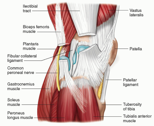
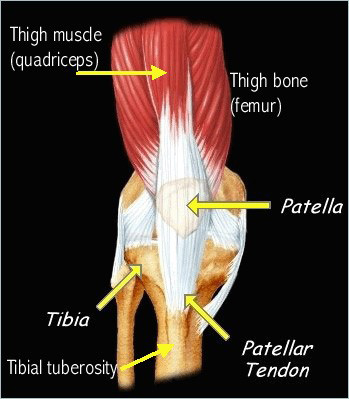
Knee Tendon Diagram : Knee Ligaments and Other Knee Stabilizers | Bone and Spine : List of skeletal muscles of the human body wikipedia.. Anatomical distribution of knee joint pain movements cartilages. Why it's a knee tendons diagram (page 1). There are two major tendons in the kneethe quadriceps and patellar. In humans and other primates, the knee joins the thigh with the leg and consists of two joints: List of skeletal muscles of the human body wikipedia.
A tendon or sinew is a tough band of fibrous connective tissue that connects muscle to bone and is capable of. This diagram depicts knee diagram tendons. Tendons are tough fibrous connective tissues that attach muscles to bones. Surgical repair of acute peroneal tendon dislocation a. Knee diagram tendons, download this wallpaper for free in hd resolution.
Brain Post: New Ligament Found in Human Knee | Could Be ... from snowbrains.com There are two major tendons in the kneethe quadriceps and patellar. The knee joint is a hinge type synovial joint, which mainly allows for flexion and extension (and a small degree of medial and lateral rotation). The knee joint is a complex structure that involves bones tendons ligaments muscles and other structures for normal function. There are several large tendons around the knee area. More collection of amazing diagrams is available in our site just look it up on the key word search. Makes up the framework of the body. Why it's a knee tendons diagram (page 1). Knee tendons medical vector illustration scheme anatomical diagram.
Many types of knee injuries can occur.
Aspect from the popliteal ligament 38. Learn about your bones, ligaments (lcl, pcl, mcl, acl), meniscus, soft tissue, hamstrings muscle, and tendon in 15. Tendon vs ligament medlineplus medical encyclopedia image. Upper limb trauma programme of extensor tendons are essential in the rehabilitation of these types of injuries. List of skeletal muscles of the human body wikipedia. Tendon, tissue that attaches a muscle to other body parts, usually bones. Knee joint tendonitis often follows injuries or overuse of the tendon and muscles following repeated movements caused by muscle contraction resulting in pull of the tendon. The knee joint is a hinge type synovial joint, which mainly allows for flexion and extension (and a small degree of medial and lateral rotation). Common knee injuries orthoinfo aaos. Diagram of the anatomy of the knee. Diagram of tendons in hand stock illustration. Muscles, tendons, ligaments, and cartilage can be strained and sprained. There are several large tendons around the knee area.
Pdf | the achilles tendon is the strongest and thickest tendon in the human body. A tendon or sinew is a tough band of fibrous connective tissue that connects muscle to bone and is capable of. Tendon, tissue that attaches a muscle to other body parts, usually bones. Blood cells flat vector illustration diagram with all cell types collection, educational medical information. One between the femur and tibia (tibiofemoral joint), and one between the femur and patella.
Knee | Radiology Key from radiologykey.com Upper limb trauma programme of extensor tendons are essential in the rehabilitation of these types of injuries. The knee joint is a complex structure that involves bones tendons ligaments muscles and other structures for normal function. Knee tendons medical vector illustration scheme, anatomical diagram. Makes up the framework of the body. Common knee injuries orthoinfo aaos. Posted on january 21, 2015 by admin. Knee diagram tendons was posted in may 29, 2015 at 4:57 pm. Tendon vs ligament medlineplus medical encyclopedia image.
There are several large tendons around the knee area.
It is formed by articulations between the patella, femur and tibia. Knee tendons medical vector illustration scheme, anatomical diagram. Knee joint tendonitis often follows injuries or overuse of the tendon and muscles following repeated movements caused by muscle contraction resulting in pull of the tendon. Implantable neuroprostheses for restoring function, 2015. Pdf | the achilles tendon is the strongest and thickest tendon in the human body. Many types of knee injuries can occur. The knee tendons are thick cords that attach the bone to muscles. The knee joint is a complex structure that involves bones tendons ligaments muscles and other structures for normal function. The knee joint is a hinge type synovial joint, which mainly allows for flexion and extension (and a small degree of medial and lateral rotation). Tendon diagram simple / leg muscle and tendon diagram google search ankle anatomy foot anatomy ankle finding a free sequence diagram tool? Tendons are tough fibrous connective tissues that attach muscles to bones. In humans and other primates, the knee joins the thigh with the leg and consists of two joints: Anatomical distribution of knee joint pain movements cartilages.
Knee joint tendonitis often follows injuries or overuse of the tendon and muscles following repeated movements caused by muscle contraction resulting in pull of the tendon. More collection of amazing diagrams is available in our site just look it up on the key word search. Anatomical distribution of knee joint pain movements cartilages. Tendons attach the knee muscles to the bone. List of skeletal muscles of the human body wikipedia.
Osgood-Schlatter Disease | Johns Hopkins Medicine from www.hopkinsmedicine.org Tendons are tough fibrous connective tissues that attach muscles to bones. Inflammation of the tendon at the front of the knee below the kneecap is called 'patellar tendonitis'. Diagram of the anatomy of the knee. There are two major tendons in the kneethe quadriceps and patellar. How the knee works dr george nicola. Below you can see a detailed diagram of the knee. 19 photos of the knee tendon anatomy diagram and name chart. Implantable neuroprostheses for restoring function, 2015.
Knee tendon diagram manual e books.
Aspect from the popliteal ligament 38. Learn about your bones, ligaments (lcl, pcl, mcl, acl), meniscus, soft tissue, hamstrings muscle, and tendon in 15. Knee joint anatomy and structures. Knee tendons medical vector illustration scheme anatomical diagram. Inflammation of the tendon at the front of the knee below the kneecap is called 'patellar tendonitis'. Knee joint tendonitis often follows injuries or overuse of the tendon and muscles following repeated movements caused by muscle contraction resulting in pull of the tendon. Posted on january 21, 2015 by admin. Posted on january 21, 2015 by admin. Knee tendon diagram manual e books. Tendons attach the knee muscles to the bone. Surgical repair of acute peroneal tendon dislocation a. It is formed by articulations between the patella, femur and tibia. Many types of knee injuries can occur.
How the knee works dr george nicola tendon diagram. Diagram of tendons in hand stock illustration.
